|
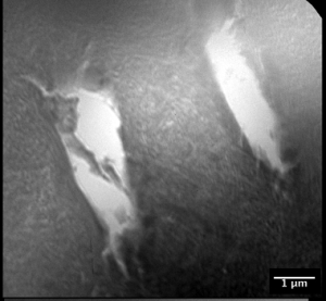
X-ray
microscopy image of dentin sample recorded at
l=2.4nm
(516.6eV) with a MZP (Drn=40nm) |
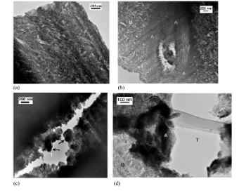
In comparison transmission
electron micrographs of transparent dentin thinned using
FIB-milling showing (a) intertubular dentin, (b) a
partially filled tubule, (c) a tubule along its long
axis partially filled with large mineral crystals, (d)
the same sample as (a)-(c) prepared with ultramicrotome
instead of FIB. (T= tubule, A= intratubular mineral, B=
intertubular mineral) (courtesy J.W. Ager, LBNL (2006)) |
|
In
Situ Studies of the Electromigration in Cu Interconnects
Recent
experiments at XM-1 demonstrated that a full-field transmission
x-ray microscope, operating at 1.8 keV photon energies,
allows detection of passivated interconnects made from
Cu or AlCu as well as vias utilizing the high material
contrast from different elements in such integrated
circuits with a resolution of about 40 nm. The mass
flow caused by electromigration in a passivated Cu interconnect
was studied in-situ at current densities up to 107
A/cm2.
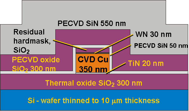
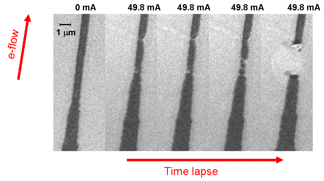
G. Schneider, M.A.
Meyer, G. Denbeaux, et al., Journal of Vacuum Science
and Technology B, 20, 3089 (2002)
|
