| |
|
|
|
|
Modern magnetic materials with applications to
magnetic storage and sensor technologies are currently focussing on
thin films and multilayered systems often accompanied with a lateral
micropatterning. Imaging magnetic microscopic processes on a
sub-micrometer length and sub-ns time scale provides key information
that will contribute significantly to a thorough understanding of the
underlying physics and will support current technological developments.
Magnetic transmission soft X-ray microscopy offers
a superior combination of the following features which match ideally
the needs both for fundamental studies in magnetism and to characterize
technologically relevant magnetic systems
-
high lateral resolution (Fresnel zone plate optical
elements)
-
sub-ns time resolution (pulsed time structure of
Synchrotron radiation)
-
elemental specificity (XMCD contrast)
-
high sensitivity to thin layers (large magnetic
absorption cross section)
-
magnetisation reversal studies (recording images in
applied magnetic fields)
-
large field of view (typical tens of microns)
-
short exposure times (typical secs per image)
|
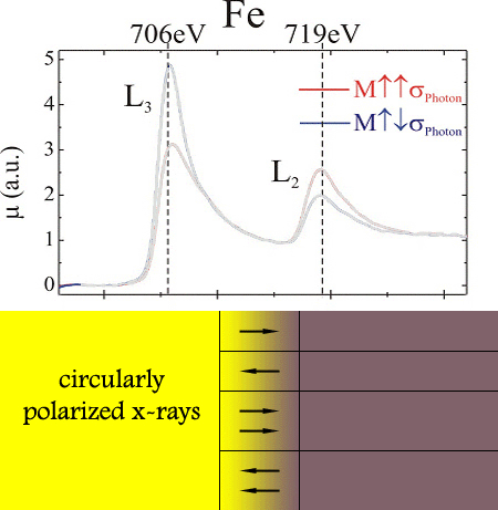
Magnetic transmission X-ray microscopy (MTXM) uses X-ray magnetic circular dichroism as
magnetic contrast mechanism. In the vicinity of element-specific
binding energies of inner core levels, such as 2p3/2
and 2p1/2 levels which correspond to L3
and L2 absorption edges, the X-ray absorption
coefficient depends strongly on the relative orientation between the
helicity of the photons and the projection of the local magnetization
onto the photon propagation direction.
With phase sensitive X-ray optics, also magnetic
phase contrast imaging has been démonstrated recently
Illuminate a ferromagnetic specimen with circularly
polarized X-rays at a specific wavelength and record the transmitted
photons with a high resolution soft X-ray microscope.
|
|
|
|
|
Examples of Recent
Results
Time Resolved Imaging of Resonant Vortex Core
Motion
The
vortex core motion in a 1.5mm permalloy disk was resonantly excited by
by spin current pulses. Analyzing the gyration radius from X-ray images
taken with 70ps time and 25nm spatial resolution the polarization of
currents characterizing the strength of the spin torque effect was
determined.
S. Kasai, et al., Phys
Rev Lett 107,
237203 (2008)
In
collaboration with U Kyoto, U Chofu and U Osaka, Japan
|
|
|
|
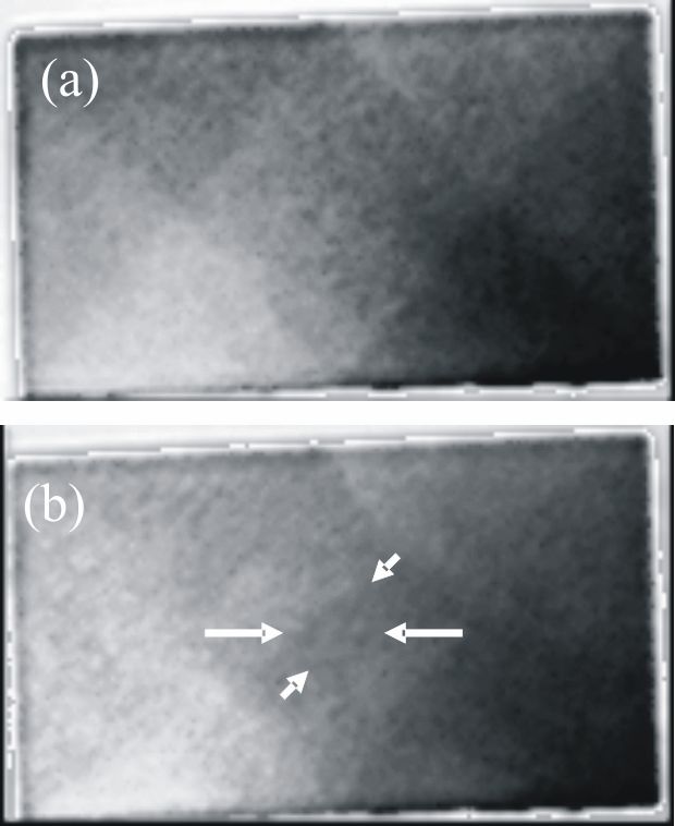
Direct
Imaging of Stochastic Current Induced Domain-Wall Motion
Pulses of
nanosecond duration and of high current density up
to 1.0×10¹² A/m2
are used to move and to deform the domain
wall. The current pulse drives the wall either undisturbed,
i.e., as composite particle through the wire, or causes structural
changes of the magnetization. Repetitive pulse measurements reveal the
stochastic nature of
current-induced domain-wall motion.
G. Meier,
et al.,
Phys. Rev. Lett. 98,
187202 (2007), also selected for Physical Review Focus
In
collaboration with
 and and 
|
|
|
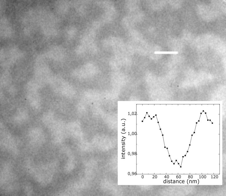
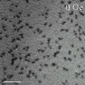
| Imaging at fundamental magnetic length scales
With a spatial resolution
of 15nm magnetic soft X-ray microscopy can probe local hysteresis
behaviour on a granular length scale in a 50 nm thick (Co83Cr17)87Pt13
nanogranular alloy film recorded at the Co L3
absorption edge (777eV). Inset: Intensity profile across a magnetic
domain (white line) proofing 15nm spatial resolution.
D.-H. Kim,
et al.
J. Appl. Phys. 99, 08H303 (2006) also selected in
Virtual
Journal of Nanoscale Science & Technology, 13(17) May 1, 2006
In collaboration with 
|
|
|
|
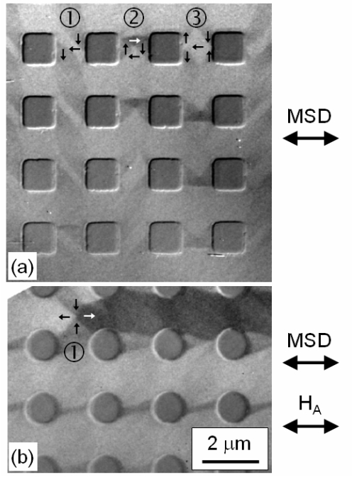
Cobalt
antidot arrays
Co
antidot arrays with 2 mm period were fabricated on X-ray transparent
membranes and imaged with MTXM:
(a)
as-grown flux closure states in array with square holes: S-state at
position 1, Landau state at position 2, flower state at position 3
(b) a
domain chain forms on application of a magnetic field with the end of
the chain comprising four 90º walls.
L. Heyderman et al., J. Magn.
Magn. Mat 316 99 (2007)
In
collaboration with
 at at

|
|
|
|
|
|
|
Problems?
Contact the webmaster.
|
DHTML Menu / JavaScript Menu
- Created Using NavStudio (OpenCube Inc.)
|