|
The
majority of biological x-ray microscopy studies are
done in the water window, which is for photon energies
between the K shell absorption edges of Oxygen (543
eV, 2.3 nm) and Carbon (284 eV, 4.4 nm). For x-ray energies
just below the Oxygen edge (e.g., 517 eV, 2.4 nm), the
absorption of mostly carbon-containing organic material
is about an order of magnitude less than the absorption
of water, permitting a natural contrast. The penetration
depth of these soft x-rays is also ideally suited to
image intact cells with a thickness of a few microns.
The photoelectric absorption, which provides contrast
in soft x-ray microscopy, is also responsible for significant
radiation damage to biological samples However, it is
possible to prepare samples in different stages of development
and then make conclusions based on statistical methods;
a common method used with electron microscopy.
Cells which are sensitive to radiation damage can be
chemically fixed to maintain cell structure during x-ray
imaging. In addition, a labeling technique for localizing
specific proteins within cellular structure can be used.
Natural antibodies are utilized to attach dense silver
and gold particles to the protein of interest. A computerized
process is used after the imaging to locate the sharp
increases in intensity to identify the regions of labeling.
For other types of experiments, we have a sample holder
for imaging cryogenically frozen cells, which mitigates
the effects of radiation damage. Because of this, chemical
fixation is not needed, which results in images with
remarkable detail.
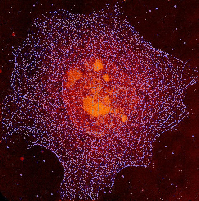
Tubulin
Network in Epithelial Cell
W. Meyer-Ilse, A. Nair/ CXRO
C. Larabell, S. Lelièvre, D. Hamamoto, M. Bissell /
Life Sciences Division
 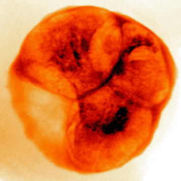 
X-ray
images of malaria infected blood cells obtained at 2.4nm
wavelength.
Left: uninfected cell, Center: newly infected cell,
Right: cell 36h after infection.
C. Magowan, W. Meyer-Ilse and J.Brown,
LBNL
Recording
a series of images of a specimen mounted in a rotational
stage with the rotation axis perpendicular to the photon
beam direction opens the avenue for X-ray
tomography at high lateral resolution to study 3-dim
structures in cells. A new X-ray microscope is currently
being set-up by the National
Center for X-ray tomography at the ALS.
|